Diagram Of Hip.and Back.muscles - Hip and thigh muscles: Anatomy and functions | Kenhub / Learn the iliopsoas, gluteal and hip adductors with diagrams now at kenhub.
byAdmin•
0
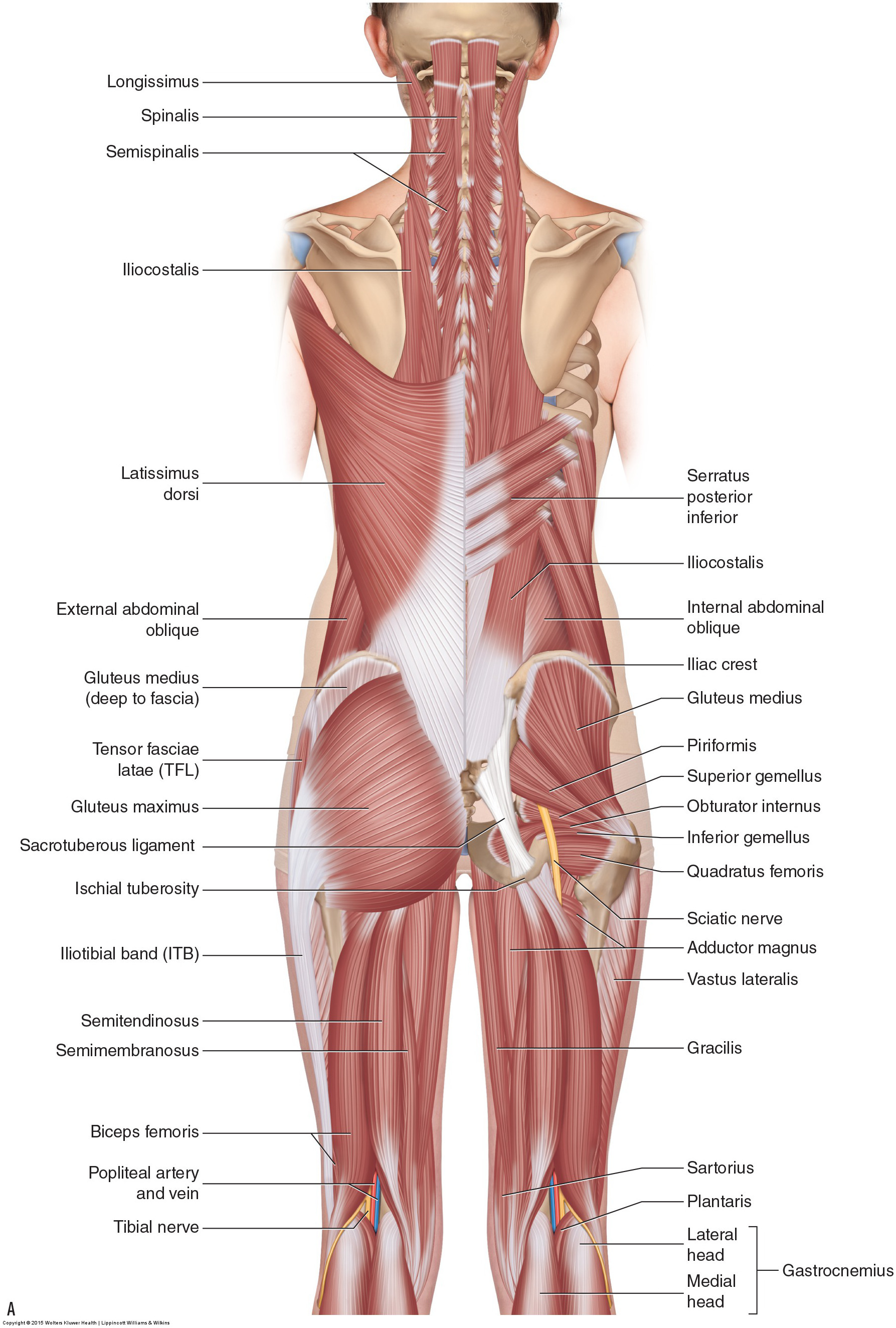
Diagram Of Hip.and Back.muscles - Hip and thigh muscles: Anatomy and functions | Kenhub / Learn the iliopsoas, gluteal and hip adductors with diagrams now at kenhub.. The red lines show where the tendons attach the muscles to the bones. The levator ani muscle along with a second muscle forms the pelvic floor. The back's muscles start at the top of the back (named the cervical vertebrae) and go to the tailbone (also named the coccyx). Key facts about hip muscles. Iliacus, psoas major, and psoas minor main function:
Learn the iliopsoas, gluteal and hip adductors with diagrams now at kenhub. The image below shows the bones from the back side of the hand. Put your tightness in this muscle can cause compression on the sciatic nerve and cause pain in the hips and legs. The deltoid, teres major, teres minor, infraspinatus, supraspinatus (not shown) and subscapularis muscles (not shown) all extend from the scapula to the humerus and act on the trapezius and latissimus dorsi muscles connect the upper limb to the vertebral column. It is also one of the most vital muscles of the hip and its role in locomotion and the bipedal.
semitendinosus Archives - Learn Muscles from learnmuscles.com Sit on the floor with your legs extended straight in front of you 2. Deadlift muscles will include knee, hip, and back extensors, which primarily include the quads, glutes, and spinal erectors. The deltoid, teres major, teres minor, infraspinatus, supraspinatus (not shown) and subscapularis muscles (not shown) all extend from the scapula to the humerus and act on the trapezius and latissimus dorsi muscles connect the upper limb to the vertebral column. Dislocation of the hip joint. All of your back muscles. Anatomy of the body hip muscles anatomy muscular system anatomy. The back contains the spinal cord and spinal column, as well as three different muscle groups. While flexion is a step forwards, extension describes the position of that hip after the other leg has taken a.
Now that you watched the video, you.
Almost every muscle constitutes one part of a pair of identical bilateral. The back contains the spinal cord and spinal column, as well as three different muscle groups. Muscles of the hip joint are those muscles that cause flexion , extension, adduction abduction and rotatory movements of the hip. Diagram of muscles and anatomy charts. Anatomy back anatomy bones gross anatomy human body anatomy muscle anatomy lower back muscles anatomy shoulder anatomy muscle diagram anatomy images. Anatomy of the body hip muscles anatomy muscular system anatomy. Sit on the floor with your legs extended straight in front of you 2. Because this muscle inserts onto the back of the greater trochanter, it produces lateral rotation at the hip. Abducts and rotates thigh laterally, flexes knee at hip, originates at the anterior superior iliac spine and inserts on the medial surface of proximal tibia. Other muscles are small and cover much less space. Dislocation of the hip joint. The back's muscles start at the top of the back (named the cervical vertebrae) and go to the tailbone (also named the coccyx). There are anterior muscles diagrams and posterior muscles diagrams.
Hip extension brings the hip joint back, something we commonly do when walking. Muscles of the back can be divided into superficial, intermediate, and deep group.since the all the back muscles originate in embryo (fetus) form by locations other than the back, muscles in the. Lower back muscles below the shoulder blade. Other muscles are small and cover much less space. Related posts of muscles of the lower back and hip diagram muscle anatomy posterior.
Running Injury Prevention Corner: Hip Adductors - DOCTORS ... from s-media-cache-ak0.pinimg.com It also covers some common conditions and injuries that can affect the. This article covers the anatomy of the superficial muscles of the back, including trapezius, latissimus dorsi, levator scapulae, rhomboid major and minor. Hip muscles and tendons march 19 2019 by luqman. Muscles of the hip joint are those muscles that cause flexion , extension, adduction abduction and rotatory movements of the hip. The red lines show where the tendons attach the muscles to the bones. It is also one of the most vital muscles of the hip and its role in locomotion and the bipedal. All of your back muscles. Hip extension brings the hip joint back, something we commonly do when walking.
Common hip and back pain causes include injury to muscles from overuse disc injurydegeneration or spinal stenosis.
Human muscle system, the muscles of the human body that work the skeletal system, that are under voluntary control, and that are concerned with movement, posture, and balance. In human anatomy, the muscles of the hip joint are those muscles that cause movement in the hip. The red lines show where the tendons attach the muscles to the bones. Diagram of muscles and anatomy charts. Muscles of the hip & thigh (quadriceps, hips). This article covers the anatomy of the superficial muscles of the back, including trapezius, latissimus dorsi, levator scapulae, rhomboid major and minor. Anatomy of the body hip muscles anatomy muscular system anatomy. There are anterior muscles diagrams and posterior muscles diagrams. Anatomy back anatomy bones gross anatomy human body anatomy muscle anatomy lower back muscles anatomy shoulder anatomy muscle diagram anatomy images. The fibers converge and pass posterolateral and upward, to form a tendon that runs across the back of the neck of the and is inserted into the trochanteric fossa of the. Luckily you've found this page to help you. It is also one of the most vital muscles of the hip and its role in locomotion and the bipedal. Some of these muscles are quite large and cover broad areas.
The former two groups, superficial and intermediate, are referred to as the extrinsic back muscles. Bend your right leg 3. Common hip and back pain causes include injury to muscles from overuse disc injurydegeneration or spinal stenosis. Related posts of muscles of the lower back and hip diagram muscle anatomy posterior. Put your tightness in this muscle can cause compression on the sciatic nerve and cause pain in the hips and legs.
Diagram Of Back Muscles - Wiring Diagram from o.quizlet.com Hip muscles and tendons march 19 2019 by luqman. Muscles of the deep back, adbominal wall, and pelv… The back's muscles start at the top of the back (named the cervical vertebrae) and go to the tailbone (also named the coccyx). Luckily you've found this page to help you. There are around 650 skeletal muscles within the typical human body. Other muscles are small and cover much less space. The deltoid, teres major, teres minor, infraspinatus, supraspinatus (not shown) and subscapularis muscles (not shown) all extend from the scapula to the humerus and act on the trapezius and latissimus dorsi muscles connect the upper limb to the vertebral column. Related posts of muscles of the lower back and hip diagram muscle anatomy posterior.
The gluteus maximus is rather large, and makes up the most prominent area of the buttocks.
Muscles of the deep back, adbominal wall, and pelv… The human back extends from the buttocks to the posterior portion of the neck and shoulders. All of your back muscles. Muscles of buttock, hip and pelvis laminated anatomy chart. Many conditions and injuries can affect the back. It also covers some common conditions and injuries that can affect the. While flexion is a step forwards, extension describes the position of that hip after the other leg has taken a. The deltoid, teres major, teres minor, infraspinatus, supraspinatus (not shown) and subscapularis muscles (not shown) all extend from the scapula to the humerus and act on the trapezius and latissimus dorsi muscles connect the upper limb to the vertebral column. Now that you watched the video, you. It joins the lower limb to the pelvic girdle. Abducts and rotates thigh laterally, flexes knee at hip, originates at the anterior superior iliac spine and inserts on the medial surface of proximal tibia. Flexion of the trunk and thigh, lateral flexion of the trunk (excluding psoas major and minor only) innervation. Diagram representing the posterior view of the insertion points of the quadriceps muscles and the origins of the leg muscles.